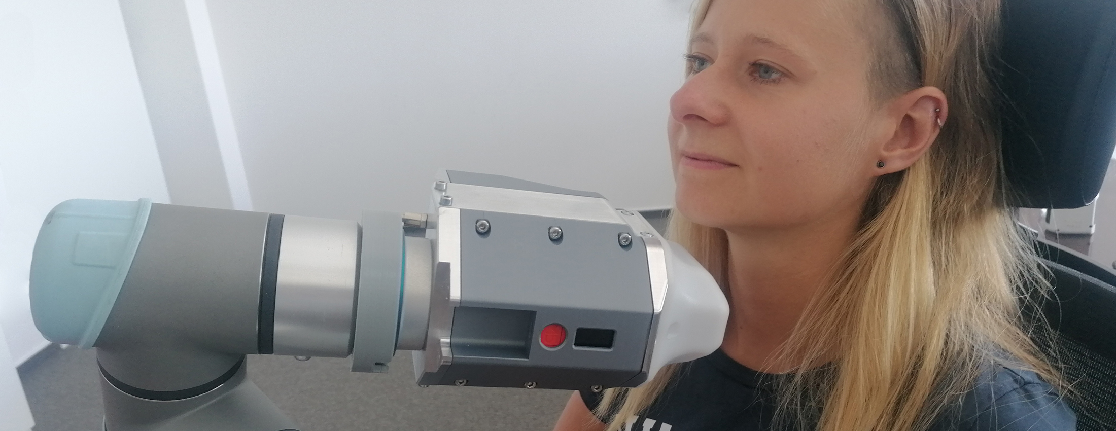


The prototype of the ThyroPIX Camera
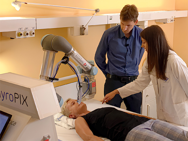
ThyroPIX - Innovative robot to help cancer patients
This video presents the prototyp of the newly developed robotic device, which could help to more accurately map the distribution of radioactive iodine in the detection and treatment of thyroid tumors. Thanks to the groundbreaking miniaturized gamma camera, capable of determining the direction of incoming radiation, it precisely locates where and how the radiopharmaceutical acts.
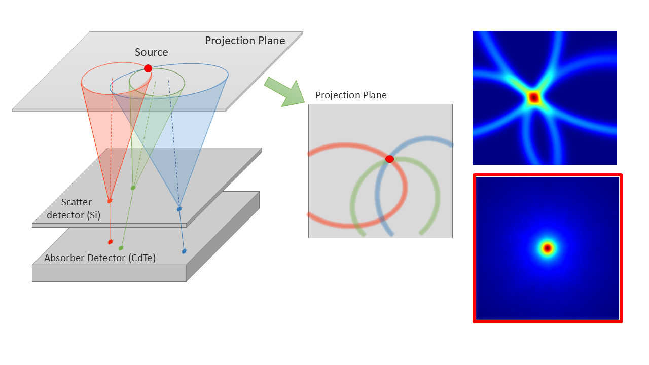
WatchCompton camera - source schema
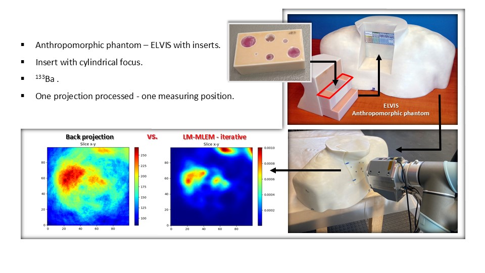
Results – Reconstruction methods experiment